![]() Figure 4 of
Le, Mol Vis 2006;
12:389-398.
Figure 4 of
Le, Mol Vis 2006;
12:389-398.
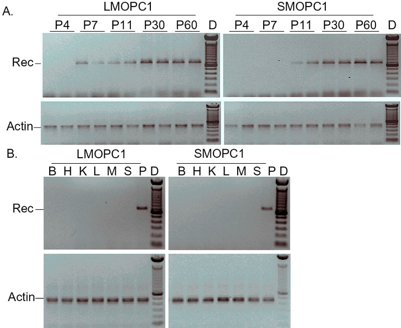
Figure 4. PCR analysis of Cre-mediated recombination
A: Time course of Cre-mediated activation of lacZ reporter gene at the DNA level using retinal DNA from P4 to P60 from the F1 double transgenic Cre/R26R mice. Retinal DNA from two different mice was used in PCR analysis. Top, inverted gel image of PCR (25 cycles) reaction diagnostic for a 710 bp Cre-mediated recombination in retinal genomic DNA of the F1 double transgenic Cre/R26R mice. Bottom, inverted gel image of PCR (20 cycles) reaction diagnostic for a 450 bp product in β-actin gene (internal DNA control). Lane D is a 100 bp DNA marker. Cre-mediated recombination was detected at P7 in the LMOPC1 mice and at P11 in the SMOPC1 mice. B: PCR analysis of ectopic Cre expression in representative tissues from both the LMOPC1 mice and the SMOPC1 mice. Lane B is brain, lane H is heart, lane K is kidney, lane L is liver, lane M is muscle, lane S is spleen; lane P is positive control DNA, and lane D is a 100 bp DNA marker. No apparent Cre-mediated recombination was identified in representative tissues from either the LMOPC1 mice or the SMOPC1 mice.