![]() Figure 2 of
Le, Mol Vis 2006;
12:389-398.
Figure 2 of
Le, Mol Vis 2006;
12:389-398.
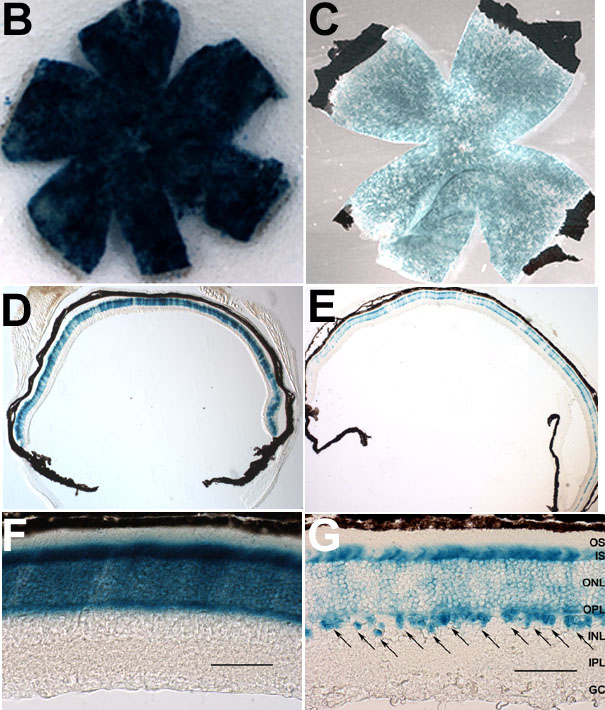
Figure 2. Localization and functional analysis of Cre expression
A: Strategy of functional assay using F1 double transgenic Cre and Cre-activatable lacZ reporter (R26R) mice. The lacZ reporter gene is expressed under the control of the generalized ROSA26 promoter after the removal of loxP-flanked transcription "STOP" sequence. Primers (e) and (f) were used to identify a 710 bp PCR product diagnostic for Cre-mediated lacZ reporter gene activation (see methods). B-G: Representative results of β-gal staining in retinal flat mounts (B,C) and sections (D-G) from six-week-old LMOPC1 mice (B,D,F) and SMOPC1 mice (C,E,G). Scale bar equals to 50 μm. Photoreceptor outer segment (OS), inner segment (IS), outer nuclear layer (ONL), outer plexiform layer (OPL), inner nuclear layer (INL), inner plexiform layer (IPL), and ganglion cell layer (GC) are labeled. Cre function was localized to the rod photoreceptors across the whole retina in both the LMOPC1 mice and the SMOPC1 mice; however, the LMOPC1 mice had a stronger Cre-activated β-gal activity. Cre function (β-gal) was also localized to the INL region (G, arrows) in the SMOPC1 mice.