![]() Figure 3 of
Demirci, Mol Vis 2006;
12:324-330.
Figure 3 of
Demirci, Mol Vis 2006;
12:324-330.
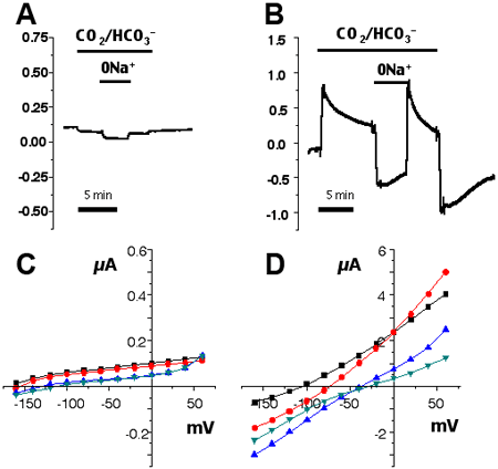
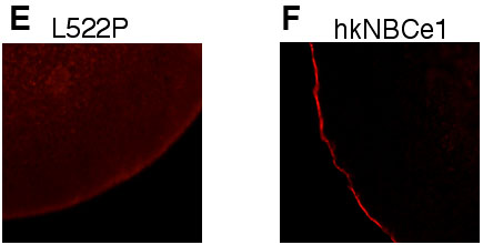
Figure 3. Functional analysis using oocytes
Two electrode voltage clamp experiments were performed on oocytes injected with L522P-hkNBCe1 cRNA (A,C,E) or wt-hkNBCe1 cRNA (B,D,F) as previously reported in the literature [7]. Oocytes were clamped at -60 mV while the bath was continuously perfused with the indicated salt solutions. HCO3- solutions were pH 7.5 at room temperature and 5% CO2 so that [HCO3-] is 33 mM. Na+ was replaced isotonically with choline. Only hkNBCe1 displays currents with HCO3- addition or Na+ removal in HCO3- solution. A,B: Currents at -60 mV. C,D: The current-voltage (IV) relationships. For IV curves, raw currents were subtracted from the baseline current measured in non-HCO3- solution (square, peak HCO3- current; circles, steady-state HCO3- current; upright triangle, peak 0Na+/HCO3- current; inverted triangles, steady-state 0Na+/HCO3- current). E,F: The results of immunostaining with a kNBCe1 specific antibody (α333) as previously described [7]. Membrane staining is obvious for wt-hkNBCe1 (F) but not for L522P (E). Water controls were negative (not shown).