![]() Figure 1 of
Demirci, Mol Vis 2006;
12:324-330.
Figure 1 of
Demirci, Mol Vis 2006;
12:324-330.
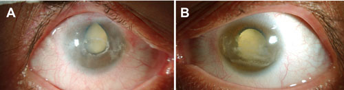
Figure 1. Appearance of the patient's anterior eye segments at age 27
Anterior segment photographs of the right (A) and left (B) eye of the patient. Both eyes demonstrate peripheral corneal vascularization and opacification, interpalpebral band keratopathy, and dense, white mature cataract. Note the loss of a distinct corneal-limbal boundary in both eyes, giving an appearance of microcornea. The right eye also shows pupil deformation (superior retraction) that seems to be due to previous glaucoma surgery.