![]() Figure 2 of
Fukushima, Mol Vis 2006;
12:310-317.
Figure 2 of
Fukushima, Mol Vis 2006;
12:310-317.
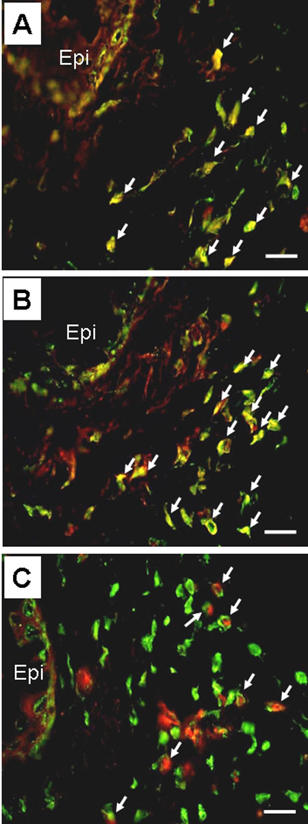
Figure 2. Immunofluorescent analyses of the cells infiltrating conjunctivas of actively immunized mice
Conjunctivas from actively immunized mice that were developing experimental allergic conjunctivitis (EC) were harvested for immunofluorescent analyses. The serial sections were first stained with FITC-labeled anti-VLA-4 antibody and then with anti-CD3 (A), anti-CD4 (B), or anti-MBP (C) antibodies, followed by development with Texas Red. Note that most of the CD3+ (A) and CD4+ (B) cells expressed VLA-4, whereas VLA-4 colocalized with MBP in only half of the eosinophils (C). Arrows indicate the cells that were stained with two different antibodies and the epithelium (Epi) is identified. The scale bars represent 20 μm. The sections shown are representative of those from five different mice.