![]() Figure 1 of
Fukushima, Mol Vis 2006;
12:310-317.
Figure 1 of
Fukushima, Mol Vis 2006;
12:310-317.
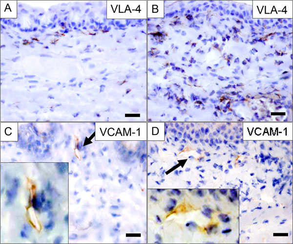
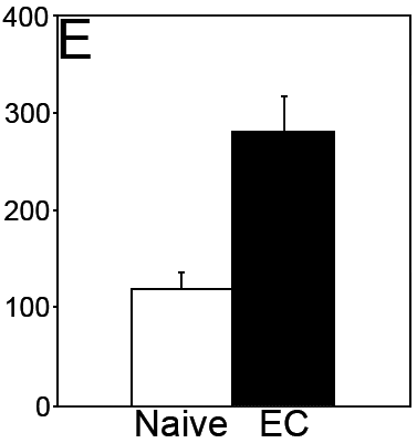
Figure 1. Expression of VLA-4 and VCAM-1 in conjunctivas of actively induced EC
Conjunctivas from naive mice (A,C) or actively immunized mice that were developing experimental allergic conjunctivitis (EC; B,D) were subjected to immunohistochemical analysis using anti-VLA-4 antibody (A,B) or anti-VCAM-1 antibody (C,D). Insets show the portions of the slides indicated by arrows at higher magnification. The scale bars in the images represent 20 μm. One representative section from five mice is shown. E: VLA-4+ cells in the lamina propria of naive and EC-developing mice were counted, and the data are presented as the number of VLA-4+ cells/mm2. Error bars represent the standard error of the mean. Note that while the number of VLA-4-expressing cells increased when EC was induced, VCAM-1 expression was not changed.