![]() Figure 7 of
Glushakova, Mol Vis 2006;
12:298-309.
Figure 7 of
Glushakova, Mol Vis 2006;
12:298-309.
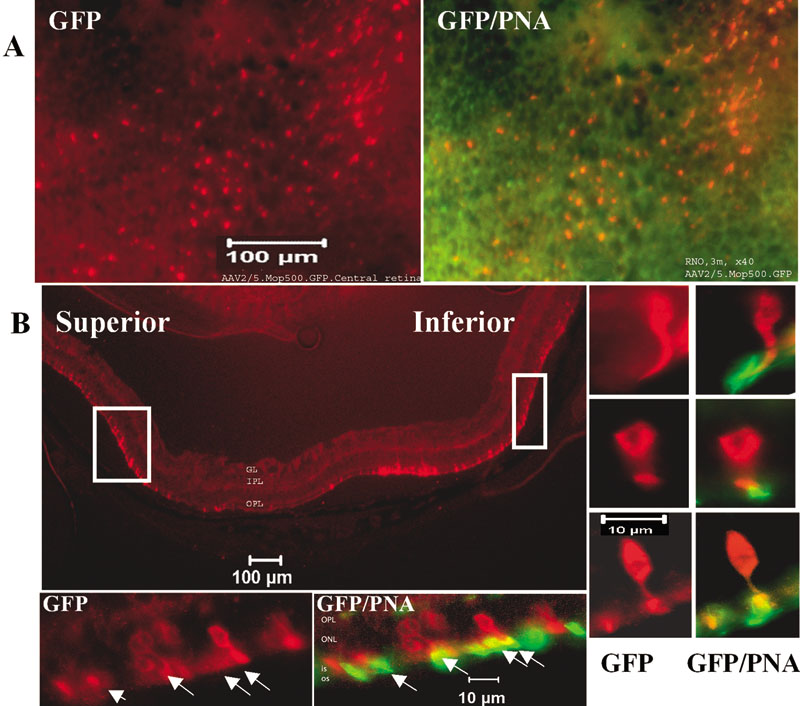
Figure 7. Green fluorescent protein expression is detected in degenerating cones of P90 rho (-/-) mouse retinas two weeks after subretinal injection of AAV2/5.mOP500.GFP
A: Fluorescent micrographs showing flatmount of central mouse retina: green fluorescent protein (GFP) expression, red emission, peanut agglutinin (PNA), green emission, and GFP/PNA, co-localization PNA-lectin and GFP-transgene expression. B: Fluorescent micrograph of vertical section. The selected fields from Inferior (right) and Superior (bottom) retina are shown at higher magnification. The ganglion layer (GL), inner plexiform layer (IPL), outer plexiform layer (OPL), outer nuclear layer (ONL), inner segments (IS), and outer segments (OS) are identified. Arrows indicate PNA and GFP co-localization.