![]() Figure 6 of
Glushakova, Mol Vis 2006;
12:298-309.
Figure 6 of
Glushakova, Mol Vis 2006;
12:298-309.
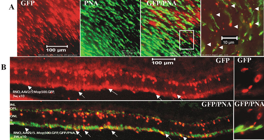
Figure 6. Green fluorescent protein expression is detected in both rods and cones of P30 rho (-/-) mouse retinas two weeks after subretinal injection of AAV2/5.mOP500.GFP
A: Fluorescent micrographs showing flatmount of central mouse retina: green fluorescent protein (GFP) expression (red emission), peanut agglutinin (PNA)-lectin binding (green emission); merge, GFP/PNA, detected on cone sheaths co-localizes with GFP. Right: A window depicts higher magnification of a selected field. B: Vertical section taken from the central retina. GFP expression (red emission); GFP/PNA co-localization of PNA-lectin binding (green emission) to cone sheaths with GFP-transgene expression. On the right, there is a selected field at higher magnification. The inner nuclear layer (INL), outer plexiform layer (OPL), outer nuclear layer (ONL), and outer segment (OS) are identified. Arrows indicate PNA-lectin detected on cone sheaths co-localizes with GFP. All images are oriented with the sclera toward the bottom and the vitreous toward the top. Scale bars are indicated for each magnification.