![]() Figure 4 of
Glushakova, Mol Vis 2006;
12:298-309.
Figure 4 of
Glushakova, Mol Vis 2006;
12:298-309.
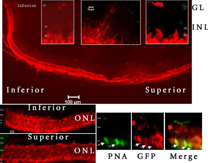
Figure 4. Green fluorescent protein expression persists in the Sprague Dawley rat retina eight months after subretinal injection of AAV2/5.mOP500.GFP
Fluorescent micrographs of frozen retinal section are presented. Green fluorescent protein (GFP) expression (red emission) is predominantly localized to the outer nuclear layer (ONL). The windows of increase magnifications on the top present superior, central and inferior fields of inner retina, and on the bottom, inferior and superior fields of outer retina. The selected field from central retina presents the co-localization (merge) of all cones sheathes (PNA; green emission) and GFP-transgene expression (GFP, red emission) at increased magnification. The arrows indicate PNA-lectin detected on cone sheaths co-localizes with GFP. The ganglion layer (GL), inner plexiform layer (IPL), inner nuclear layer (INL), outer plexiform (OPL), outer nuclear layer (ONL), outer segment (OS), and inner segment (IS) are identified.