![]() Figure 3 of
Glushakova, Mol Vis 2006;
12:298-309.
Figure 3 of
Glushakova, Mol Vis 2006;
12:298-309.
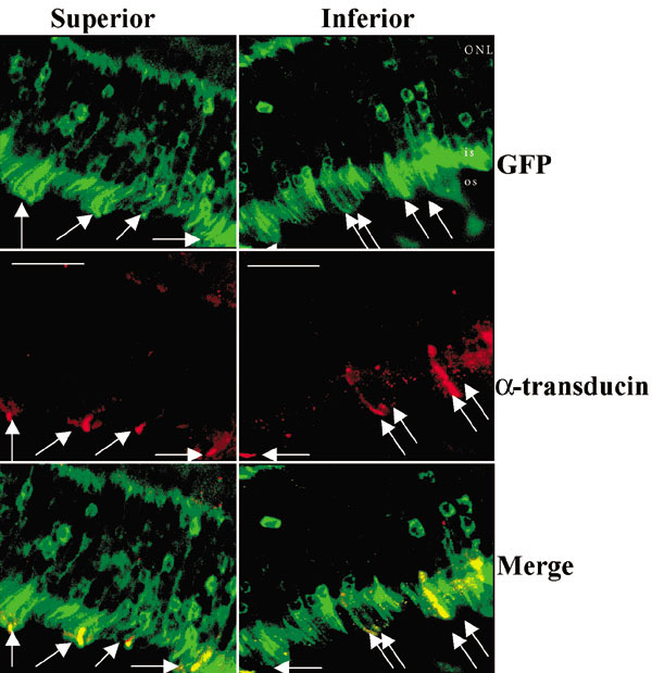
Figure 3. Patterns of cone transduction by AAV2/5.mOP500.GFP vector in treated rat eye: two weeks after sub-retinal injection
Fluorescent micrographs of retinal superior (Superior) and inferior (Inferior) fields are presented. Co-localization (Merge) of cone specific α-transducin (α-transducin) and GFP-transgene expression (GFP) in ONL are pointed by arrows. All cones revealed by α-transducin labeling from either inferior, or superior field are GFP-positive. The scale bar represents 30 μm.