![]() Figure 2 of
Glushakova, Mol Vis 2006;
12:298-309.
Figure 2 of
Glushakova, Mol Vis 2006;
12:298-309.
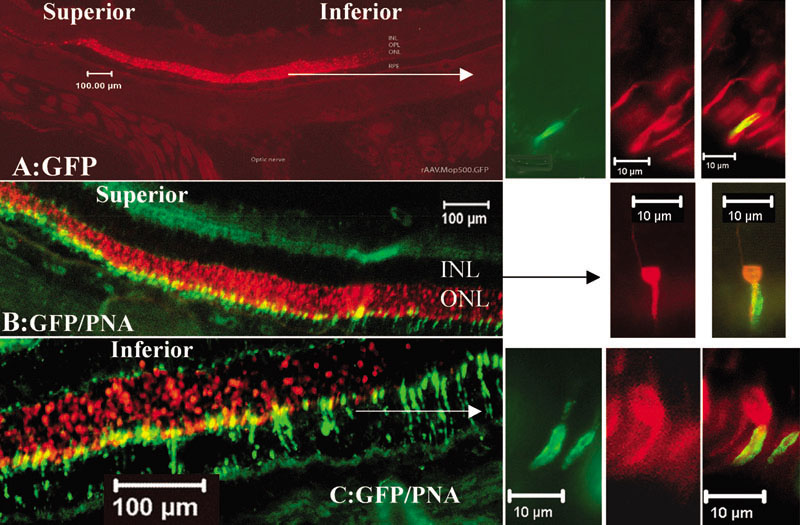
Figure 2. Green fluorescent protein expression is detected in both rods and cones of the Sprague Dawley rat retina two weeks after sub-retinal injection of AAV2/5.mOP500.GFP
Fluorescent micrographs of vertical sections from treated rat eye are presented. GFP expression (A; red emission) is exclusively localized to the outer nuclerr layer (ONL). Superior (B) and inferior (C) retinal fields are shown at increased magnification. Peanut agglutinin (green emission) detected on cone sheaths co-localizes with green fluorescent protein (GFP, red emission). On the right, selected central (A), superior (B), and inferior (C) retinal fields are shown at higher magnifications. All images are oriented with the sclera toward the bottom and the vitreous toward the top. The inner nuclear layer (INL) is identified.