![]() Figure 6 of
Flynn, Mol Vis 2006;
12:271-282.
Figure 6 of
Flynn, Mol Vis 2006;
12:271-282.
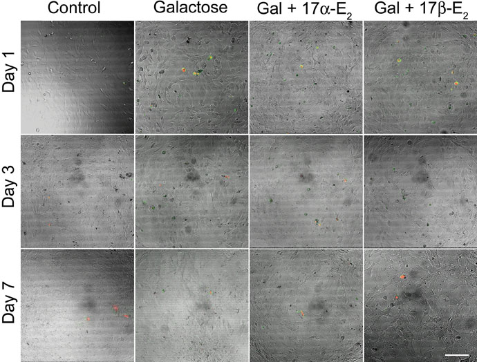
Figure 6. Determination of cell viability of BLECs through 7 days of Gal exposure as assessed by annexin V-propidium iodide labeling
Random representative confocal images of bovine lens epithelial cells stained with both annexin V (green) and propidium Iodide (red) as described in methods. The images indicate minimal positive staining with either annexin V (indicates apoptosis) or propidium iodide (indicates necrosis) for all experimental treatments through seven days. The scale bar represents 20 μm.