![]() Figure 2 of
Flynn, Mol Vis 2006;
12:271-282.
Figure 2 of
Flynn, Mol Vis 2006;
12:271-282.
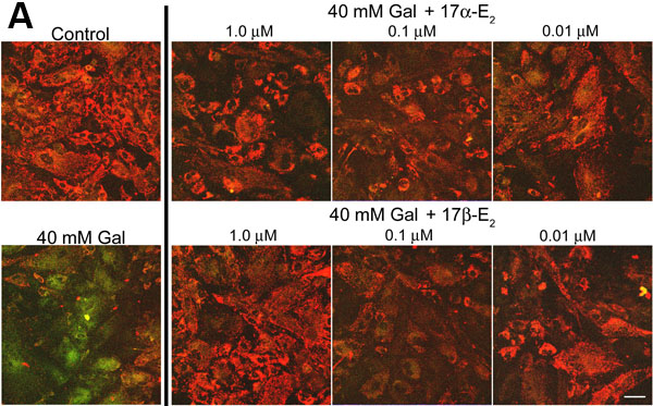
Figure 2. 17α-estradiol and 17β-estradiol prevent mitochondrial membrane depolarization with galactose-treated bovine lens epithelial cells
A: Representative confocal images of galactose (Gal)-exposed BLECs supplemented with a pharmacological dose (1 μm) to a physiological dose (0.01 μm) of estradiol. Far left panels show the loss of mitochondrial membrane potential in 3 day Gal-treated cells as compared to control. The set of panels on the right illustrate estradiol's protection across the entire dose range for both estrogen stereoisomers. The scale bar represents 20 μm. B: Statistical analyses of confocal images. All treatments with the exception of the control cells, received either Gal or Gal supplemented with a given dose of estradiol. Gal treatment alone resulted in significant depolarization (asterisk; n=8, p>0.05). 17α-estradiol and 17β-estradiol significantly prevented (+) depolarization across the entire range of doses (n=8, p>0.05). Error bars represent the standard error of the mean.