![]() Figure 6 of
Kuszak, Mol Vis 2006;
12:251-270.
Figure 6 of
Kuszak, Mol Vis 2006;
12:251-270.
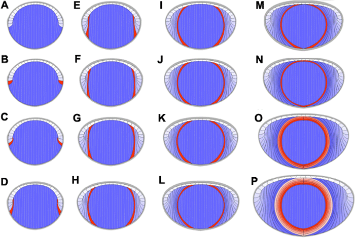
Figure 6. Correct interpretation of secondary fiber development
This animation begins with a qualitative rendering of a generic adult lens radial cell column face of a lens just after completion of the primary fiber cell mass or embryonic nucleus (A). By highlighting a nascent pair of developing fibers (orange) the onset of and general scheme of lifelong lens growth can be appreciated (B-P). Developing fibers simultaneously rotate about their long axis, migrate posteriorly, and elongate until they are oriented essentially parallel to the lens polar axis (B-E). Note the constancy of the position of the highlighted pair of developing fibers for the remainder of the animation (F-P). This is a dynamic demonstration of the end of developing fiber rotation and migration. The rest of fiber elongation takes place independent of rotation and migration. Note the lens dimensions are enlarging due to more fibers developing at the periphery (light blue to blue).
Note that the slide bar at the bottom of the quicktime movie can be used to manually control the flow of the movie. If you are unable to view the movie, a representative frame is included below.
| This animation requires Quicktime 6 or later. Quicktime is available as a free download. |
