![]() Figure 5 of
Kuszak, Mol Vis 2006;
12:251-270.
Figure 5 of
Kuszak, Mol Vis 2006;
12:251-270.
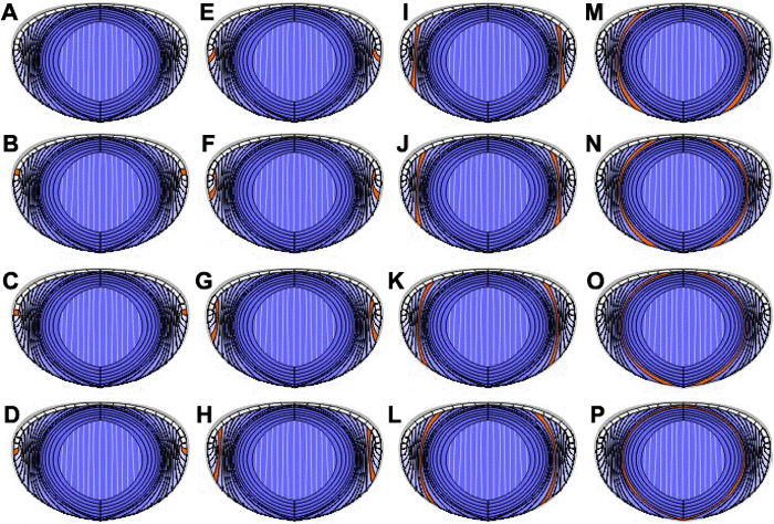
Figure 5. Incorrect interpretation of secondary fiber development
The animation begins with a qualitative rendering of a generic adult lens radial cell column face (A). It is this type of drawing, based on light and scanning electron microscopic analysis of lenses from a variety of species, that has led to the notion that fibers simultaneously, rotate, migrate and elongate until they reach their sutural destinations. Indeed, if a pair of nascent differentiating fibers is highlighted (orange) in this image (B) and then presumably one step further along in their development in successive copies of the same image (C-P), then animating these images on a timeline, would appear to support the above notion. However, this interpretation is incorrect because it does not take into account the fact that lenses become larger as they grow.
Note that the slide bar at the bottom of the quicktime movie can be used to manually control the flow of the movie. If you are unable to view the movie, a representative frame is included below.
| This animation requires Quicktime 6 or later. Quicktime is available as a free download. |