![]() Figure 12 of
Kuszak, Mol Vis 2006;
12:251-270.
Figure 12 of
Kuszak, Mol Vis 2006;
12:251-270.
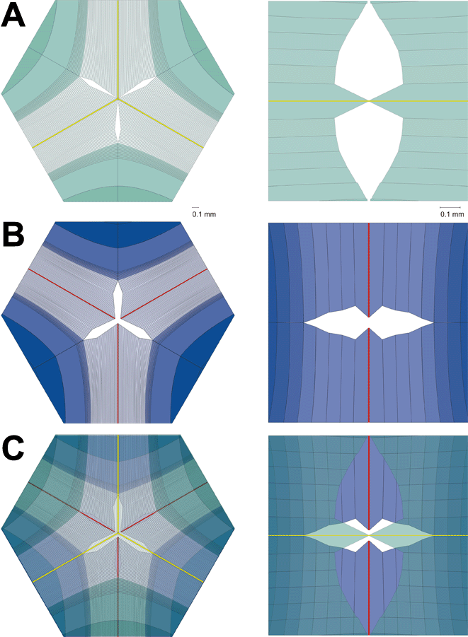
Figure 12. A Comparison of Y and line suture distal end segment formation
Comparison of percent of fiber specialized as anterior (A), middle, or posterior segment (B), and posterior polar caps (C), respectively. Note that there is a proportionately greater amount of distal end segmentation in line as compared to Y suture lenses that is directly related to the number of suture branches or by extrapolation, a broader range of intrashell fiber length.
