![]() Figure 4 of
Nallathambi, Mol Vis 2006;
12:236-242.
Figure 4 of
Nallathambi, Mol Vis 2006;
12:236-242.
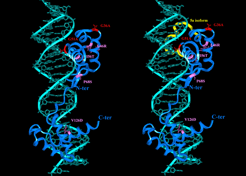
Figure 4. Overview of PAX6 paired domain DNA complex modeled structure
The PAX6 mutated structure and the PAX6 5a isoform (yellow) are shown as a ribbon model with the mutated residues from other studies (pink) depicted as sticks. The mutations identified in this study are shown in red. The DNA bases (light green) and the ribbon representation of the PAX 6 protein are represented in blue.
