![]() Figure 3 of
Nallathambi, Mol Vis 2006;
12:236-242.
Figure 3 of
Nallathambi, Mol Vis 2006;
12:236-242.
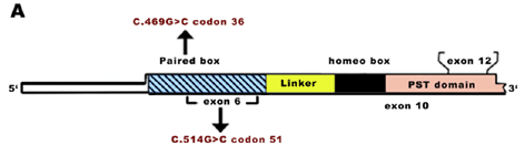
Figure 3. Schematic of PAX6 and amino acid conservation analysis of PAX6 protein
A: A schematic of PAX6 shows the location of nucleotide changes in the paired box that were found in this study. B: Amino acid sequence alignment of the human PAX6 paired domain (amino acids 34-57) homologs were compared with other species (PAX6_DROME is the Drosophila Pax-6 eyeless homolog and PAX6_toy is the toy-twin of eyeless homolog). The Gly36 and Gly51 shown in red are highly conserved.
B:
               36                                         51
               *                                          *
PAX6_HUMAN   HSGVNQLGGVFNGRPLPDSTRQKIVELAHSGARPCDISRILQVSNGCVSKIL
PAX6_XENLA   HSGVNQLGGVFNGRPLPDSTRQKIVELAHSGARPCDISRILQVSNGCVSKIL
PAX6_MOUSE   HSGVNQLGGVFNGRPLPDSTRQKIVELAHSGARPCDISRILQVSNGCVSKIL
PAX6_DROME   HSGVNQLGGVFGGRPLPDSTRQKIVELAHSGARPCDISRILQVSNGCVSKIL
PAX6_toy     HSGINQLGGVYNGRPLPDSTRQKIVELAHSGARPCDISRILQVSNGCVSKIL
PAX6_RAT     HSGVNQLGGVFNGRPLPDSTRQKIVELAHSGARPCDISRILQVSNGCVSKIL
PAX6_COTJA   HSGVNQLGGVFNGRPLPDSTRQKIVELAHSGARPCDISRILQVSNGCVSKIL
PAX6_BRARE   HSGVNQLGGVFNGRPLPDSTRQKIVELAHSGARPCDISRILQVSNGCVSKIL
|