![]() Figure 1 of
Nallathambi, Mol Vis 2006;
12:236-242.
Figure 1 of
Nallathambi, Mol Vis 2006;
12:236-242.
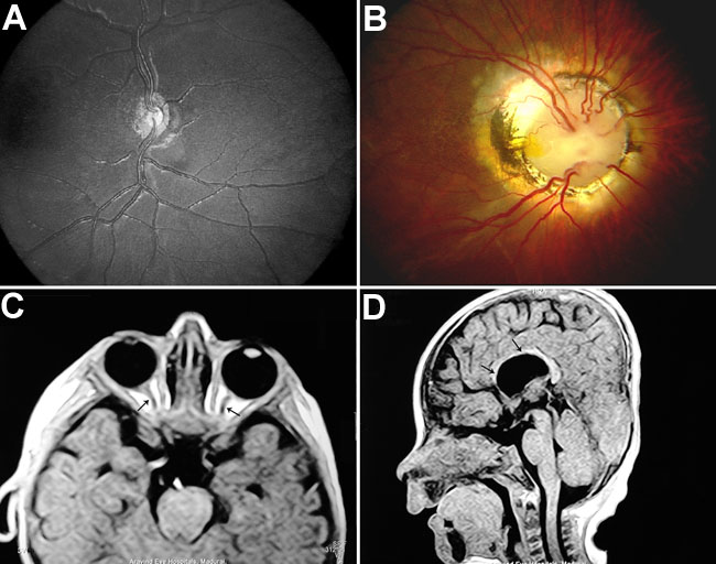
Figure 1. Fundus photography and MRI scan report of the affected probands
A: Ophthalmoscopic appearance of a 4-year-old male showing an abnormally small optic nerve head that is surrounded by a yellowish mottled peripapillary halo boarded by a dark pigment ring (double ring sign). B: Ophthalmic exam of a 6-year-old boy showed the fundus picture of optic disc coloboma with large wafer-like defect, enlarged disc funnel shaped excavation surrounded by an elevated annulus of chorioretinal pigmentary disturbance. Blood vessels emerge from the rim of excavation like the spokes of a wheel. C: MRI scan of axial view (T1 weighted image [WI]) shows thinning of optic nerves in proband ONH 4-1 (arrow). D: MRI scan (saggital view, T1 WI) shows the congenital absence of septum pellucidum with hypogenesis of corpus callosum (arrow).