![]() Figure 8 of
Mullins, Mol Vis 2006;
12:224-235.
Figure 8 of
Mullins, Mol Vis 2006;
12:224-235.
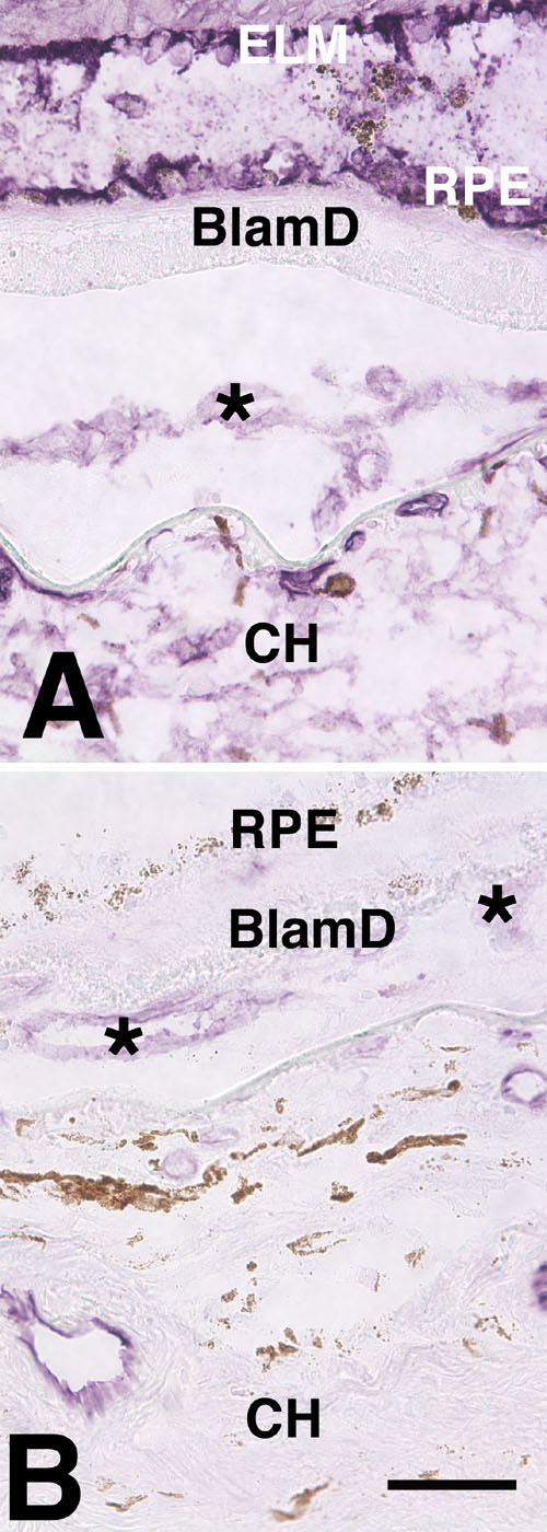
Figure 8. Comparison of ICAM-1 and ICAM-2 eyes with CNV
A: In eyes with choroidal neovascular membranes, ICAM-1 exhibits weak labeling throughout the dystrophic retina and in vascular elements within the choroidal neovascular membrane (asterisk). B: ICAM-2 is restricted to vascular elements (asterisks). The external limiting membrane (ELM), basal laminar deposits (BlamD), and choroid (CH) are identified. Scale bar represents 50 μm.