![]() Figure 4 of
Mullins, Mol Vis 2006;
12:224-235.
Figure 4 of
Mullins, Mol Vis 2006;
12:224-235.
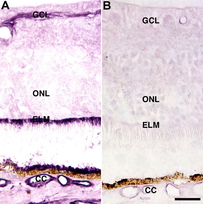
Figure 4. Comparison of ICAM-1 and ICAM-2 labeling in a healthy eye
A: ICAM-1 immunoreactivity is widespread, with labeling observed in the ganglion cell layer (GCL), external limiting membrane (ELM), RPE, choriocapillaris (CC), and other choroidal vessels [30]. B: ICAM-2 is restricted in its distribution to retinal and choroidal vasculature. The outer nuclear layer (ONL) is also identified. Scale bar represents 25 μm.