![]() Figure 5 of
Chen, Mol Vis 2006;
12:196-204.
Figure 5 of
Chen, Mol Vis 2006;
12:196-204.
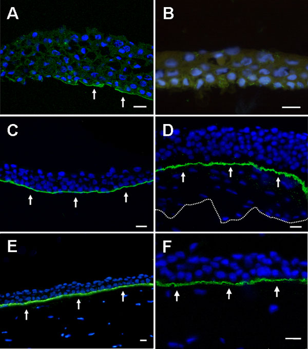
Figure 5. Collagen VII in the epithelial sheets from eyes with recurrent corneal erosion
Immunostaining for collagen VII resulted in two reference patterns of response in epithelial sheets from the eyes with recurrent corneal erosion. These responses were either a discontinuous line of positive staining along the basal aspect (A, arrows) or no staining at all (B). Nuclei were counterstained blue with 4',6'-diamidino-2-phenylindole. C: A continuous linear pattern for the staining of collagen VII (arrows) at the basement membrane complex was noted in mechanically separated normal epithelial sheet, indicating that the plane of cleavage is deeper than the collagen VII layer in the traumatized corneal epithelium of normal eye. D: A uniform staining of the basement membrane complex for collagen VII was observed as a continuous line (arrows) in the partially lamellar keratectomized corneal tissue, with the above epithelium and underlying stroma counterstained blue. The dotted line represents the inferior contour of lamellar keratectomy sample. E: A continuous linear pattern for the staining of collagen VII (arrows) in the basement membrane complex was discernible in the normal corneoscleral rim. F: A picture taken at a higher magnification and limited to a single plane by confocal microscopy in the normal corneoscleral rim also revealed a continuous linear pattern for the collagen VII staining (arrows) in the basement membrane complex. The green-stained anchoring fibril layer seemed to be slightly undulating under such a high magnification. The scale bar represents 20 μm. A,C,D,F: Confocal scanning micrographs. B,E: Light micrographs.