![]() Figure 4 of
Chen, Mol Vis 2006;
12:196-204.
Figure 4 of
Chen, Mol Vis 2006;
12:196-204.
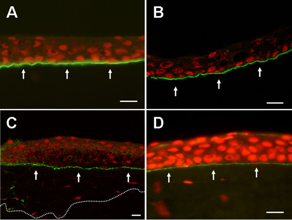
Figure 4. Laminin 5 in the epithelial sheets from the eyes with recurrent corneal erosion
A: Staining for laminin 5 showed a line of positive response along the base of recurrent corneal erosion epithelial sheet (nuclei counterstained red with propidium iodine). B: linear expression of laminin 5 was along the basal side of mechanically separated normal epithelium (arrows). C: Staining for laminin 5 also had a positive response and manifested as a linear expression along the basal aspect of epithelium in partially lamellar keratectomized corneal tissue (arrows). The dotted line represents the inferior contour of lamellar keratectomy sample. D: Immunofluorescence staining for laminin 5 exhibited a linear pattern (arrows) at the interface of epithelium and stroma in the corneoscleral rim. The scale bar represents 20 μm. A,D: Light micrographs. B,C: Confocal scanning micrographs.