![]() Figure 3 of
Chen, Mol Vis 2006;
12:196-204.
Figure 3 of
Chen, Mol Vis 2006;
12:196-204.
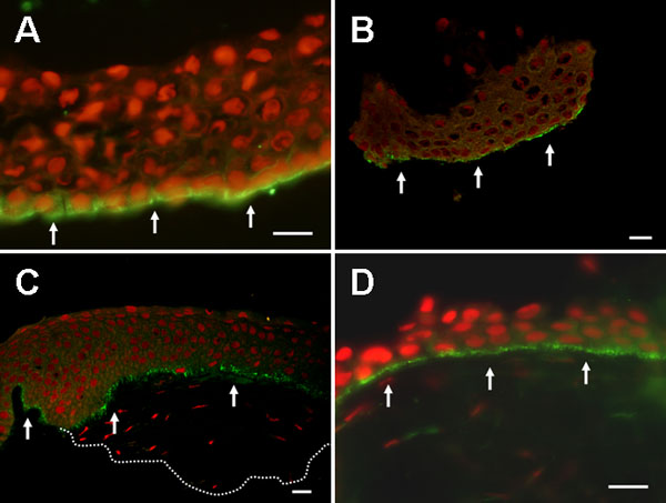
Figure 3. Integrin β4 in the epithelial sheets from the eyes with recurrent corneal erosion
A: Staining for integrin β4 was positive with a linear manifestation (arrows) and localized only at basal epithelial cells of recurrent corneal erosion epithelial sheet (nuclei counterstained red with propidium iodine). B: Staining for integrin β4 demonstrated a typical punctate pattern in a linear fashion (arrows), consistent with hemidesmosome arrangement in mechanically separated normal epithelial sheets. C: Integrin β4 was deposited in a punctate pattern in basal epithelial cells (arrows) throughout the whole length of sample in partially lamellar keratectomized corneal tissue. The dotted line represents the inferior contour of lamellar keratectomy sample. D: A punctate pattern of integrin β4 was observed in a linear expression at the basal epithelial layer as well in adhesion complex of the corneoscleral rim (arrows). The scale bar represents 20 μm. A,D: Light micrographs. B,C: Confocal scanning micrographs.