![]() Figure 2 of
Chen, Mol Vis 2006;
12:196-204.
Figure 2 of
Chen, Mol Vis 2006;
12:196-204.
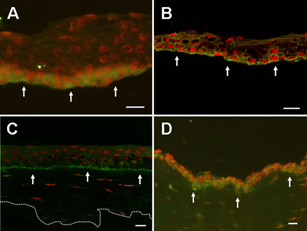
Figure 2. Integrin β1 in epithelial sheets from eyes with recurrent corneal erosion
A: Integrin β1 immunostaining of the nonadherent corneal epithelium in recurrent corneal erosion (RCE) showed strongly positive results (green) at the basal and suprabasal layers of detached epithelial sheet (arrows; nuclei counterstained red with propidium iodine). B: A similar pattern of integrin β1 was observed in mechanically separated normal epithelial sheets, showing positive pericellular staining at suprabasal and basal layers (arrows). C: Integrin β1 immunofluorescence showed positive pericellular staining mainly at basal layer in partially lamellar keratectomized corneal tissue (arrows). The dotted line represents the inferior contour of lamellar keratectomy sample. D: Integrin β1 immunofluorescence staining is observed at suprabasal and basal cell layers of adhesion complex in corneoscleral rim (arrows). The scale bar represents 20 μm. A,D: Light micrographs. B,C: Confocal scanning micrographs.