![]() Figure 1 of
Chen, Mol Vis 2006;
12:196-204.
Figure 1 of
Chen, Mol Vis 2006;
12:196-204.
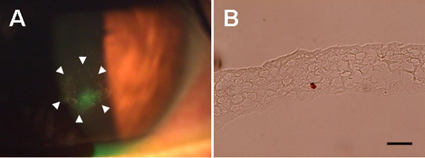
Figure 1. Clinical picture and micrograph of recurrent corneal erosion
A: A flank epithelial defect was stained green with sodium fluorescein. An area of loosened epithelium surrounding the flank epithelial defect was outlined with arrow heads. Biopsy of recurrent corneal erosion (RCE) was performed in this area. B: Light micrograph of RCE sample. An epithelial sheet comprising 4-7 layers of cells without apparent stromal components was noted. The scale bar represents 20 μm.