![]() Figure 2 of
Zhao, Mol Vis 2006;
12:15-25.
Figure 2 of
Zhao, Mol Vis 2006;
12:15-25.
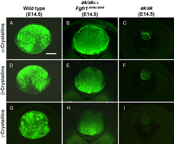
Figure 2. Analysis of crystallin gene expression in lenses derived from Fgfr1Δtmk/Δtmk ES cells
Expression of α-crystallins (A-C), β-crystallins (D-F), and γ-crystallins (G-I) were analyzed by immunofluorescence with specific antibodies. The expression of these lens structural proteins in rescued lenses from ak/ak<->Fgfr1Δtmk/Δtmk chimeras (B,E,H) are shown together with those of wild-type (A,D,G) and ak/ak (C,F,I) control lenses. Lenses derived from Fgfr1Δtmk/Δtmk ES cells expressed these crystallin genes at levels comparable to those of wild-type lenses. Notice that α-crystallins were expressed in both the lens epithelial and fiber cells, whereas β- and γ-crystallin expression was only detected in the lens fiber cells. In ak/ak lenses, α- and β-crystallins were expressed at very low level and no γ-crystallin expression was observed. The scale bar represents 100 μm.