![]() Figure 1 of
Zhao, Mol Vis 2006;
12:15-25.
Figure 1 of
Zhao, Mol Vis 2006;
12:15-25.
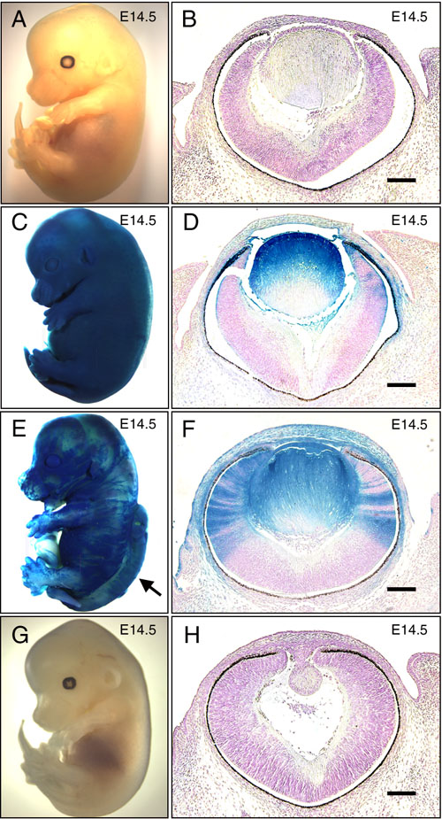
Figure 1. Histological analysis of ak/ak<->Fgfr1Δtmk/Δtmk chimeras
ES cell-derived tissues were identified by β-galactosidase expression. X-gal staining was performed on wild-type (A,B), ak/ak<->Fgfr1Δtmk/+ (C,D), ak/ak<->Fgfr1Δtmk/Δtmk (E,F) and ak/ak (G,H) embryos at E14.5. Both whole mount pictures (A,C,E,G) and embryonic eye sections (B,D,F,H) are shown. The presence of rescued lens that stained positive for X-gal in ak/ak<->Fgfr1Δtmk/Δtmk (F) demonstrates that Fgfr1 deficient ES cells are capable of forming lenses with normal morphology. A neural tube defect in the ak/ak<->Fgfr1Δtmk/Δtmk chimera (E) is indicated by an arrow. In the ES-cell derived lenses (D,F), the lack of penetration of the X-gal staining reagents in whole mount embryos prevented the staining of the deep lens fibers. The scale bars represent 100 μm in B,D,F,H.