![]() Figure 9 of
Boatright, Mol Vis 2006;
12:1706-1714.
Figure 9 of
Boatright, Mol Vis 2006;
12:1706-1714.
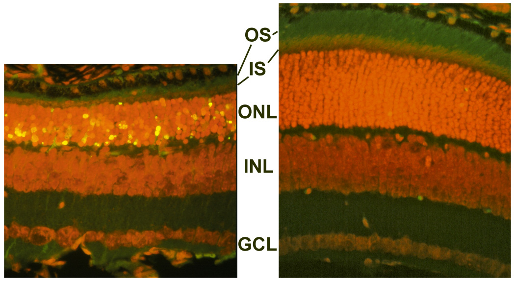
Figure 9. Effect of TUDCA on rd10 mouse retinal morphology and TUNEL: Further observation
Fluorescence microscopy using a B-2A longpass emission fluorescence filter allows further observation of the preservation of photoreceptor inner segments (IS) and outer segments (OS; the light-capturing structure of the cell) present in TUDCA-treated (right) versus vehicle-treated retina sections (left). Similar to confocal imagery (Figure 8), TUNEL-positive nuclei (green/yellow signal) are seen to be abundant in vehicle-treated sections, but rare in TUDCA-treated sections. TUDCA treatment provided significant preservation of photoreceptor nuclei number in the outer nuclear layer (ONL). Treatment had no discernable effect on the inner nuclear layer (INL) or ganglion cell layer (GCL).