![]() Figure 3 of
Boatright, Mol Vis 2006;
12:1706-1714.
Figure 3 of
Boatright, Mol Vis 2006;
12:1706-1714.
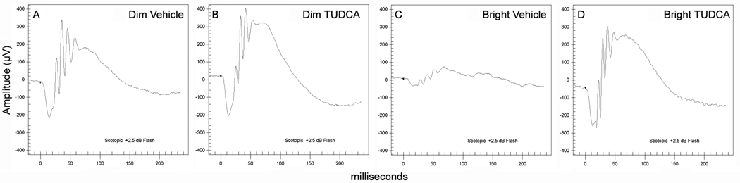
Figure 3. Effect of TUDCA on LIRD mouse retina function: Representative ERG traces at 24 h post-light exposure
To test whether TUDCA protects the retina from bright-light damage, mice were pretreated with TUDCA or vehicle only and then exposed to bright or dim light. After light treatment (24 h), electroretinograms (ERGs) were recorded as an outcome measure of retina function. Representative ERGs are shown, in response to a 137 cd sec/m2 Ganzfeld flash (+2.5 dB). A illustrates a mouse that received vehicle only and was exposed to dim light. This represents a normal ERG profile for this ERG flash stimulus. B is from a mouse treated with TUDCA and exposed to dim light. This tracing is normal, suggesting that TUDCA by itself does not affect the ERG. C was recorded from a mouse that was treated with vehicle only and exposed to bright light. This condition reflects extensive damage to the retina, and the ERG shows a greatly diminished a-wave, b-wave, and oscillatory potentials. D illustrates a trace recorded from a mouse treated with TUDCA and exposed to bright light. The ERG is very similar to the response shown in A, the vehicle treated dim-light mouse, suggesting that retina function is unimpaired. The implicit times are the same among all four groups, and the number of oscillatory potential peaks appears the same. This experiment shows that major differences are the profound loss of ERG signal (about 80% reduction) following light damage, and the apparent full protection from light damage by TUDCA.