![]() Figure 1 of
Boatright, Mol Vis 2006;
12:1706-1714.
Figure 1 of
Boatright, Mol Vis 2006;
12:1706-1714.
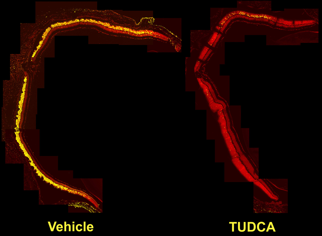
Figure 1. Effect of TUDCA on LIRD mouse retina morphology and apoptosis: 24 h post-light exposure
Composite images of confocal micrographs of LIRD mouse retina sections assayed for apoptosis by fluorescent TUNEL. The images were assembled into a composite from confocal micrographs laid over one another in Photoshop to provide a complete image of a retina section. A representative composite of the results obtained with vehicle (left) versus TUDCA (right) injections is shown. In each composite, the optic nerve is to the left and the cornea, which was removed, would be to the right. The composites represent the full diameter of the adult eye, which is approximately 3 mm. Bright light exposure induced massive apoptosis (yellow signal) and morphological damage in photoreceptor cells and retinal outer nuclear layer of vehicle-treated (left) but not TUDCA-treated eyes (right). The percentage of TUNEL-positive photoreceptors was greatly reduced in TUDCA- versus vehicle-treated LIRD mice (49.4±11% versus 11.9±4.2%; p=0.0074, t=3.443, df=9; counts per field as detailed in Methods).