![]() Figure 1 of
Lai, Mol Vis 2006;
12:1687-1691.
Figure 1 of
Lai, Mol Vis 2006;
12:1687-1691.
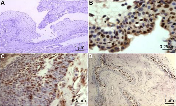
Figure 1. Representative positive and negative immunostaining for BPDE-like DNA adducts in paraffin sections of pterygium
Representative negative immunostaining is shown in A (100X), positive immunostaining in the epithelial layer is shown in both B (400X) and C (200X). Positive immunostaining in the epithelial layer and weak positive staining in the subepithelial fibrous sarcoma are shown in D (100X).