![]() Figure 5 of
Linberg, Mol Vis 2006;
12:1674-1686.
Figure 5 of
Linberg, Mol Vis 2006;
12:1674-1686.
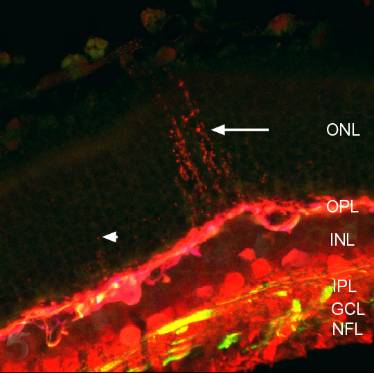
Figure 5. Triple labeled 7 day detached cat retina
A 7 day detached cat retina triple labeled with antibodies against calretinin (red), neurofilament (green) and calbindin D (blue). A large array of beaded HC outgrowths (arrow) rises more than halfway through the outer nuclear layer (ONL) while shorter arrays (arrowhead) also originate from the red, anti-calretinin-dominated processes of the distal outer plexiform layer (OPL). Anti-neurofilament-positive processes are more numerous in the inner plexiform layer (IPL) and ganglion cell layer (GCL). INL represents inner nuclear layer; NFL represents nerve fiber layer.