![]() Figure 2 of
Linberg, Mol Vis 2006;
12:1674-1686.
Figure 2 of
Linberg, Mol Vis 2006;
12:1674-1686.
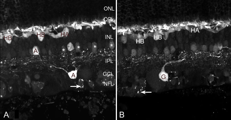
Figure 2. Confocal micrographs of the immunofluorescent labeling patterns of anti-calretinin in normal cat retina
Confocal micrographs of the immunofluorescent labeling patterns of anti-calretinin in normal cat retina. A: While the antibody to calretinin labels both types of HC (HA, HB), the B-type cell is labeled more intensely. Certain amacrine cell types (A) label brightly on both sides of the inner plexiform layer (IPL); others, notably the AII amacrine cells (*) label less robustly. A subpopulation of fibers in the nerve fiber bundles labels strongly with this antibody (arrows). B: In addition, a ganglion cell (G) is anti-calretinin-positive and sends its primary dendritic trunk into the mid-IPL. ONL represents outer nuclear layer; INL represents inner nuclear layer; GCL represents ganglion cell layer; NFL represents nerve fiber layer.