![]() Figure 12 of
Linberg, Mol Vis 2006;
12:1674-1686.
Figure 12 of
Linberg, Mol Vis 2006;
12:1674-1686.
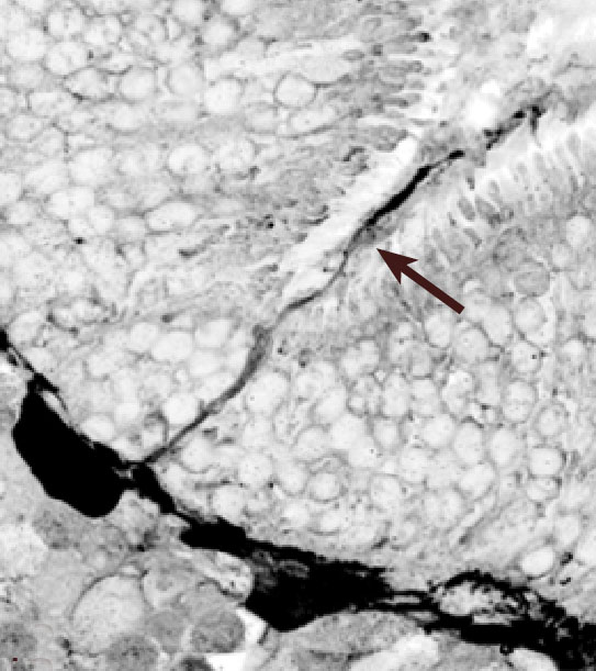
Figure 12. Confocal micrograph of a α-calretinin-positive HC outgrowth
Confocal micrograph of an α-calretinin-positive HC outgrowth (arrow) that extends across a narrowed ONL at a retinal fold in a 28 day detached cat retina. This is one in a series of 21 optical sections harvested at 0.3 μm intervals that are compiled as an AVI movie loop. Both the digitally inverted image and the AVI file were created using Image Pro Plus software.
Note that the slide bar at the bottom of the quicktime movie can be used to manually control the flow of the movie. If you are unable to view the movie, a representative frame is included below.
| This animation requires Quicktime 6 or later. Quicktime is available as a free download. |