![]() Figure 11 of
Linberg, Mol Vis 2006;
12:1674-1686.
Figure 11 of
Linberg, Mol Vis 2006;
12:1674-1686.
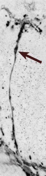
Figure 11. Confocal micrograph of an anti-calretinin-positive HC outgrowth in a section of cat retina that has been detached for 28 days
Confocal micrograph of an anti-calretinin-positive HC outgrowth in a section of cat retina that has been detached for 28 days. This process rises from the OPL and branches distally (arrow) near the retinal surface. This image is one in a series of 38 optical sections harvested at 0.3 μm intervals that are compiled as an AVI movie loop. Both the digital image inversion and the AVI file were created using Image Pro Plus software.
Note that the slide bar at the bottom of the quicktime movie can be used to manually control the flow of the movie. If you are unable to view the movie, a representative frame is included below.
| This animation requires Quicktime 6 or later. Quicktime is available as a free download. |