![]() Figure 2 of
Kannan, Mol Vis 2006;
12:1649-1659.
Figure 2 of
Kannan, Mol Vis 2006;
12:1649-1659.
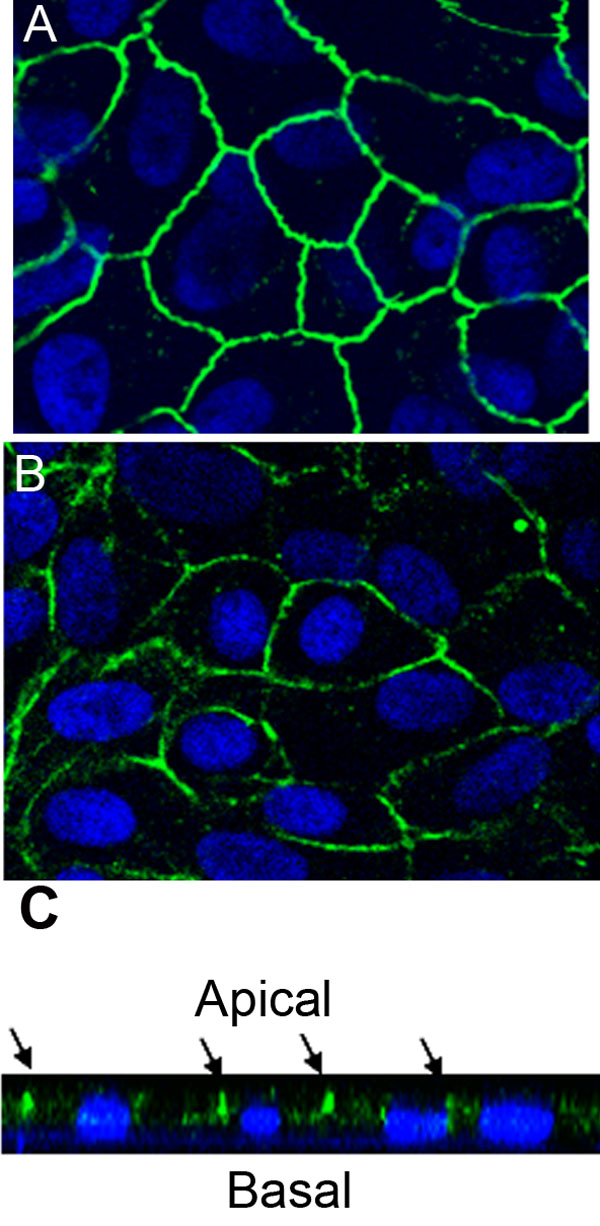
Figure 2. Evidence for tight junctions and polarity in ARPE-19 cells cultured on transwell filters for four weeks
Details of staining procedure are provided in Methods. A: Immunofluorescence of tight junctions visualized by occludin staining in ARPE-19 cells. B: Immunofluorescence with Na/K ATPase showed that it was mainly localized on the apical plasma membrane of the cells. C: Confocal vertical (X-Z) section of Na/K+ ATPase staining showing apical localization.