![]() Figure 5 of
Kim, Mol Vis 2006;
12:1640-1648.
Figure 5 of
Kim, Mol Vis 2006;
12:1640-1648.
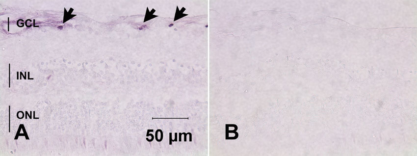
Figure 5. Immunohistochemical detection of annexin II in the normal human retina
In accordance with the observed gene expression profile, annexin immunoreactivity is primarily observed in distinct areas of the GCL (arrows) and, to a lesser degree, diffusely in the nerve fiber layer (A). No labeling was observed in the absence of the primary antibody (B).