![]() Figure 2 of
Kim, Mol Vis 2006;
12:1640-1648.
Figure 2 of
Kim, Mol Vis 2006;
12:1640-1648.
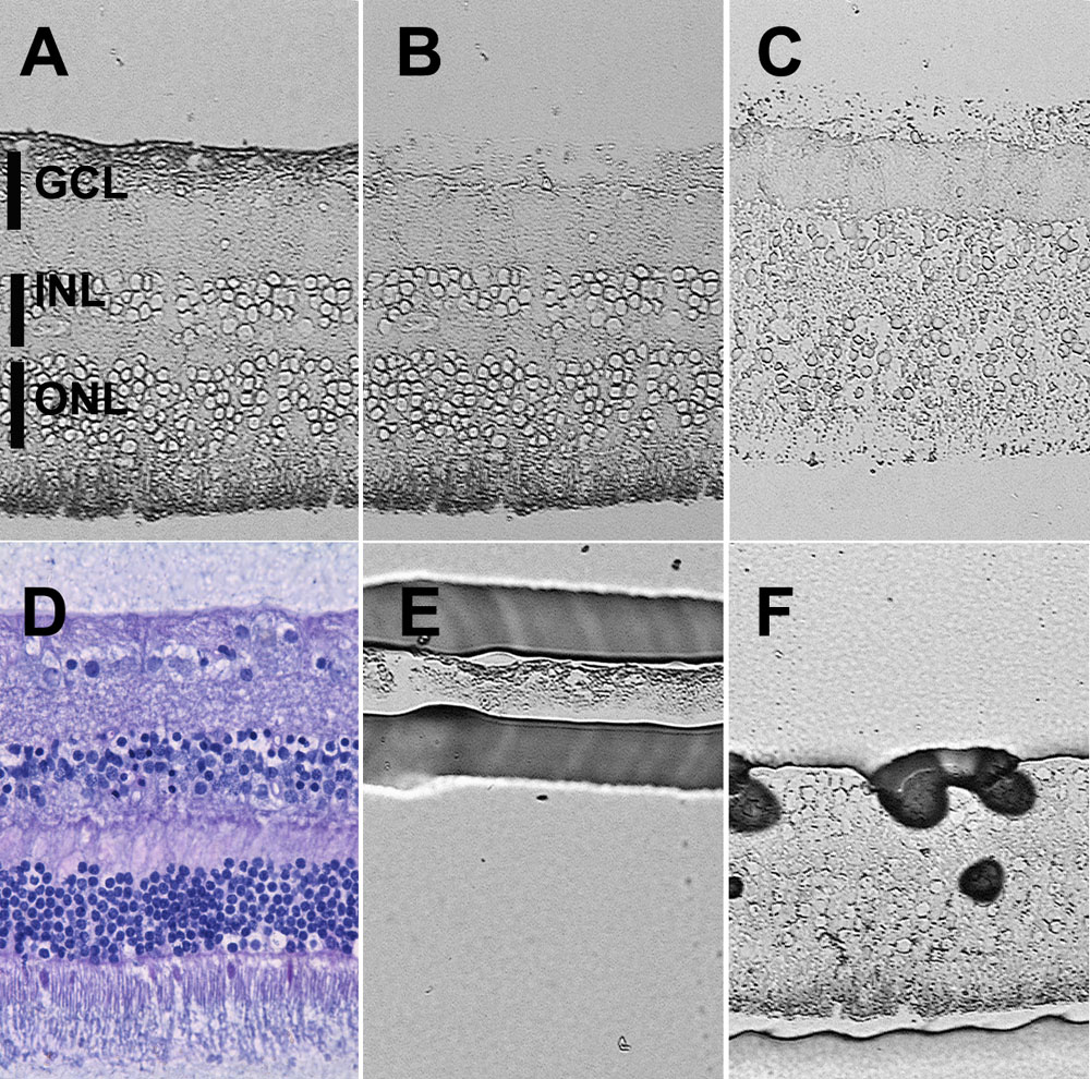
Figure 2. Laser capture microdissection of unfixed sections of human retina
Despite the reduced morphological preservation inherent in unfixed tissue, the distinct layers of the retina are clearly discernible. This series of images demonstrates that isolates of ganglion cell layer (GCL) or OR of high purity can be obtained. A: Tissue prior to capture. B: Remaining tissue after capture of the GCL, C: Remaining tissue after capture of the OR, E: isolated GCL material. F: Isolated OR material. Vertical bars mark areas collected for analyses. D: Histological appearance of adjacent portions of the retina from the same donor stained with hematoxylin and eosin. (INL represents inner nuclear layer, ONL represents outer nuclear layer).