![]() Figure 2 of
Duncan, Mol Vis 2006;
12:1632-1639.
Figure 2 of
Duncan, Mol Vis 2006;
12:1632-1639.
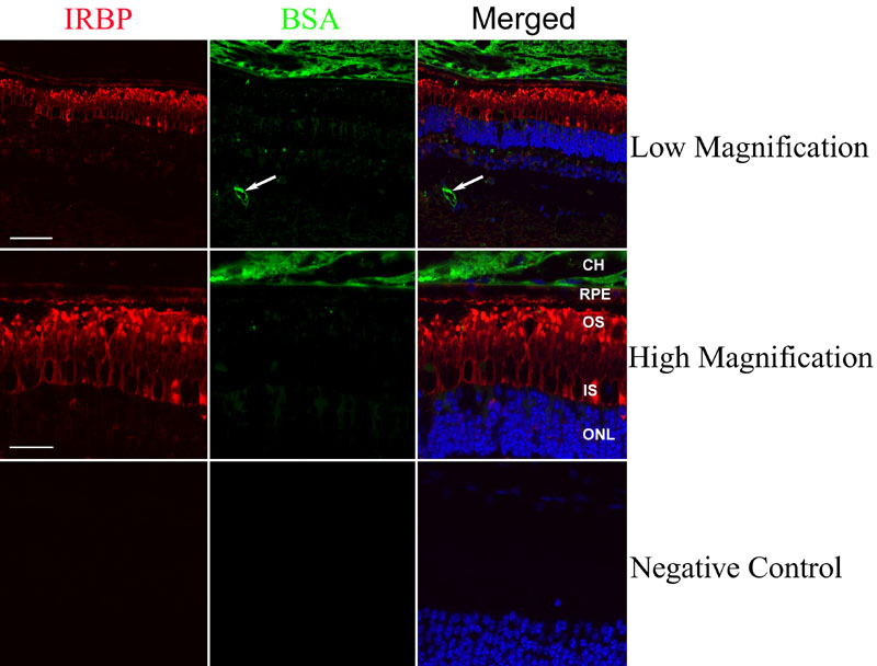
Figure 2. Confocal immunofluorescence analysis of interphotoreceptor retinoid-binding protein and bovine serum albumin in bovine retina
Immunoreactivity for interphotoreceptor retinoid-binding protein (IRBP; red fluorescence) is visible throughout the interphotoreceptor matrix (IPM). Labeling for bovie serum albumin (BSA; green fluorescence) is associated only with the lumen of retinal (arrows) and choroidal blood vessels. There is no significant labeling for BSA in the IPM, as defined by IRBP labeling (red). The dark ovals within the IPM and just above the outer nuclear layer are cone photoreceptor inner segments. Cell nuclei appear blue after DAPI staining. The following abbreviations are used: choroid (CH), retinal pigment epithelium (RPE), outer segment (OS), inner segment (IS), outer nuclear layer (ONL). Top row: Scale bar represents 75 μm; middle and bottom rows: Scale bar represents 30 μm.