![]() Figure 6 of
Srivastava, Mol Vis 2006;
12:1615-1625.
Figure 6 of
Srivastava, Mol Vis 2006;
12:1615-1625.
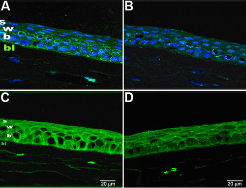
Figure 6.
Confocal images (at 63X magnification) of normal and KC corneal sections using anti-α-enolase antibody (A and B) and anti-β-actin antibody (C and D). In the images, s: superficial layer; w: wing cell layer; b:basal cell layer of the epithelium; and BAL: Bowman's layer. A and B show a relatively intense and uniform immunostaining with anti-α-enolase antibody of the epithelial basal, wing and superficial cells of the normal corneas but in the KC cornea, a maximum staining of the basal cells, which was greatly diminished in the wing and superficial cells. C and D show almost similar results as described in A and B on immunoreactivity with anti-β-actin antibody. DAPI staining of the sections is seen as blue fluorescence in the nuclei of the cells.