![]() Figure 2 of
Micera, Mol Vis 2006;
12:1594-1600.
Figure 2 of
Micera, Mol Vis 2006;
12:1594-1600.
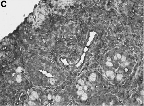
Figure 2. Tβ4 is overexpressed by VKC conjunctival biopsies
A: Total RNA from conjunctival specimens was reverse-transcribed and amplified with Tβ4 or GAPDH primers to generate PCR products by real-time PCR. Scatter plot of Tβ4 mRNA expression in VKC conjunctiva, normalized for GAPDH levels and expressed as Cts (mean±SEM), with respect to healthy conjunctiva. B: representative melting curve of Tβ4 amplification. C: Immunoreactivity for Tβ4 in VKC conjunctiva as detected by the avidin-biotin peroxidase technique. Nuclei were counterstained with hematoxylin (40X).