![]() Figure 1 of
Micera, Mol Vis 2006;
12:1594-1600.
Figure 1 of
Micera, Mol Vis 2006;
12:1594-1600.
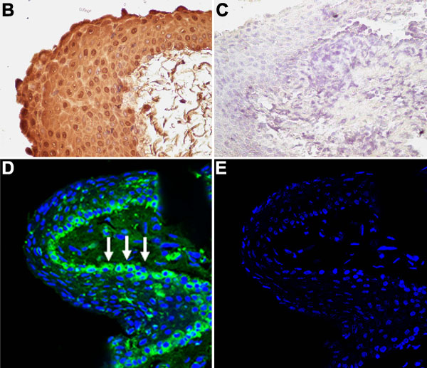
Figure 1. Tβ4 mRNA/protein is expressed by healthy conjunctival biopsies
A: Specific Tβ4 fluorescent amplification in healthy conjunctival specimens as detected by reverse-transcription of conjunctival total RNA and amplification with Tβ4 or GAPDH primers to generate PCR products by PCR analysis. B: Immunoreactivity for Tβ4 in healthy conjunctiva as detected by the avidin-biotin peroxidase technique. Nuclei were counterstained with hematoxylin (40X). D: Fluorescent Tβ4 immunoreactivity (green) tended to localize in the conjunctival epithelium, mainly in the basal membrane (white-arrows), and few Tβ4 positive cells localized in the stroma. Blue staining is referred to Toto-3 nuclear marker (60X/oil immersion). C,E: Irrelevant IgG immunoreactivity (isotype) for light and fluorescent staining, respectively (40X and 60X/oil immersion).