![]() Figure 5 of
Namekata, Mol Vis 2006;
12:1586-1593.
Figure 5 of
Namekata, Mol Vis 2006;
12:1586-1593.
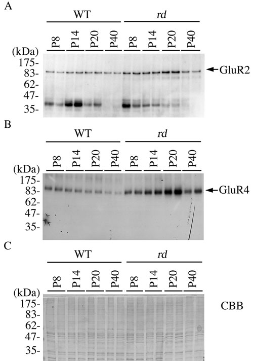
Figure 5. Immunoblot analysis of GluR2 and GluR4 in the postnatal mice retina
Retinal extracts were prepared from two wild-type and rd mice at P8, P14, P20, and P40. The extracts were separated on 8% polyacrylamide gel and analyzed by immunoblotting using anti-GluR2 (A) or -GluR4 (B) antibody. Once GluR4 was detected, the proteins fixed on the membrane were stained with CBB (C). Note the higher GluR4 protein expression in rd mice compared with wild-type mice between P14 and P40.