![]() Figure 3 of
Namekata, Mol Vis 2006;
12:1586-1593.
Figure 3 of
Namekata, Mol Vis 2006;
12:1586-1593.
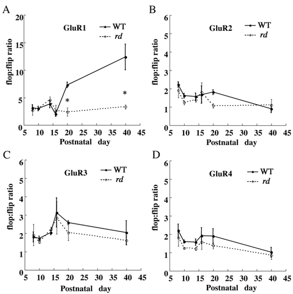
Figure 3. GluR flop:flip ratio in the postnatal mice retina
The ratio was calculated by quantitative real-time RT-PCR analysis. The flop:flip ratio in GluR1 was dramatically increased between P16 and P40 in wild-type mice (A). Note the significantly low GluR1 flop:flip ratio in rd mice as compared to wild-type mice at P20 and P40. Each data point represents the mean±SEM of the values obtained from three or four independent experiments. *p<0.05.