![]() Figure 9 of
Nickerson, Mol Vis 2006;
12:1565-1585.
Figure 9 of
Nickerson, Mol Vis 2006;
12:1565-1585.
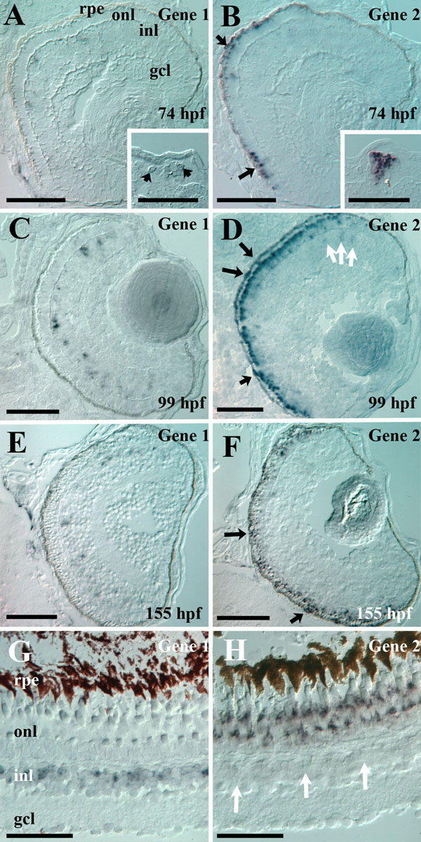
Figure 9. In situ hybridizations
Zebrafish IRBP Gene 1 and Gene 2 are differentially expressed. Retinal cryosections were obtained from 74 hpf (A, B) albb4 zebrafish embryos, 99 hpf (C, D) and 155 hpf (E, F) albb4 zebrafish larvae, and adult Tue zebrafish eyes (G, H) and were hybridized with Gene 1- (A, C, E, G) and Gene 2- (B, D, F,H) specific probes. Note that albb4 zebrafish do have low levels of pigment in the RPE; dark arrows show Gene 2 hybridization. White arrows show weak expression of Gene 2 sporadically in the INL. Insets in A and B shows Gene 1 and Gene 2 expression in developing pineal; contrast was enhanced to show weak expression of Gene 1. Scale bars represent 50 μm; rpe represent retinal pigmented epithelium, onl represent outer nuclear layer; inl represent inner nuclear layer; gcl represent ganglion cell layer.