![]() Figure 7 of
Nickerson, Mol Vis 2006;
12:1565-1585.
Figure 7 of
Nickerson, Mol Vis 2006;
12:1565-1585.
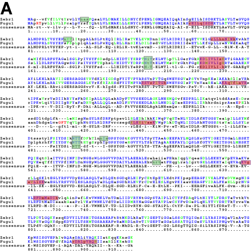
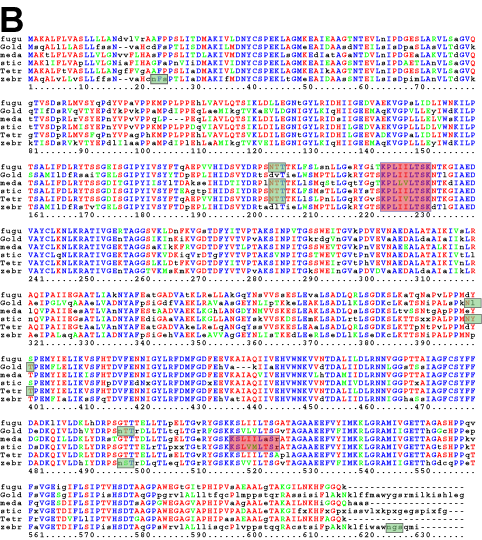
Figure 7. Multiple alignment of fish amino acid sequences
A: Gene 1 from zebrafish (zebr1) and fugu (fugu1) are aligned over Repeats 1 through 3, about 930 amino acids. B: Gene 2 Repeats 1 and 2 orthologs from fugu, goldfish, medaka, stickleback, tetraodon, and zebrafish are aligned over about 620 amino acids. Motifs identified: Signal peptide (leader sequence) cleavage sites are shown by the arrow. Glycosylation sites, NX(T|S), are shown in gray boxes with a transparent green fill. Hyaluronan binding sites, (R|K)X7(R|K), are shown in a blue box with a transparent light red fill. Identical amino acids across all aligned positions are shown in blue. Red illustrates identical residues in 3 to 5 of the 6 possible identities. Green illustrates residues with similar chemical properties at a given position.

