![]() Figure 4 of
Yoshida, Mol Vis 2006;
12:1558-1564.
Figure 4 of
Yoshida, Mol Vis 2006;
12:1558-1564.
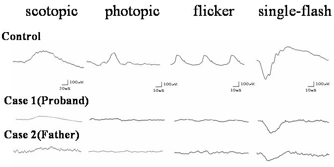
Figure 4. Electrophysiological recordings from the affected individuals and a normal subject
The scotopic rod electroretinogram (ERG), photopic cone ERG, 30-Hz flicker ERG, and bright flash rod cone mixed ERG amplitudes are markedly reduced in the proband. Those of the proband's father exhibited a similar trend, although more severely reduced.