![]() Figure 2 of
Yoshida, Mol Vis 2006;
12:1558-1564.
Figure 2 of
Yoshida, Mol Vis 2006;
12:1558-1564.
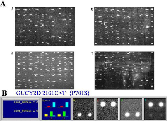
Figure 2. Leber congenital amarousis arrayed primer extension-based microarray assay hybridized to probes generated from proband's genomic DNA
A: Grayscale images for each fluorescent dideoxy nucleotide are used for the sequence analysis. B: Sequence alteration in the third base of codon 701 of the GUCY2D gene, analyzed by the GENORAMA software. Grayscale bitmaps corresponding to all four fluorescent dideoxy nucleotides at the base to be determined are shown enabling visual analysis. T signals in the sense area and A in the antisense area are indicative for a sequence alteration.