![]() Figure 3 of
Palamalai, Mol Vis 2006;
12:1543-1551.
Figure 3 of
Palamalai, Mol Vis 2006;
12:1543-1551.
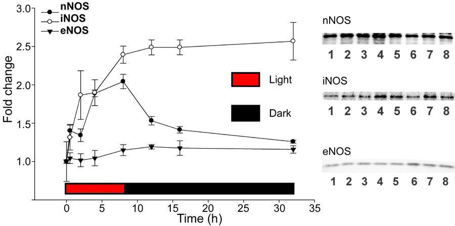
Figure 3. Western analyses of nNOS, iNOS, and eNOS
Quantification of immunoblots of anti-nNOS, anti-iNOS, and anti-eNOS antibody are shown with representative western blots. Lane 1: No light exposure; lane 2: 30 min light exposure; lane 3: 2 h light exposure; lane 4: 4 h light exposure; lane 5: 8 h light exposure; lane 6: 8 h light exposure followed by 4 h of darkness; lane 7: 8 h light exposure followed by 8 h of darkness; lane 8: 8 h light exposure followed by 24 h in darkness. The results are from three gels from one set of retinas and presented as mean±standard deviation. In each case, the fold changes were normalized to the unexposed NOS optical density (lane 1).